Back Of Skull Anatomy / Skull, posterior view with labels - Axial Skeleton Visual ... - The skull is the bony skeleton of the head.. Anatomy next provides anatomy learning tools for students and teachers. The anterior fossa is formed by the orbital plates of the frontal bone, cribriform plate of the ethmoid, and lesser wings of the sphenoid. In order to be light, the skull is made up by flat and irregular bones, and has hollow spaces called the sinuses. The skull base is the inferior portion of the neurocranium. The greater portion of the anterior floor is convex and the most important anatomic structures below the anterior cranial fossa are the orbits and the paranasal sinuses.
The skull has a single occipital condyle.7 the skull consists of five major bones: It offers protection to the brain, eye balls, inner ears, and nasal passages. The axial & appendicular skeleton. The skull bones can be classified into two groups: The anterior fossa is formed by the orbital plates of the frontal bone, cribriform plate of the ethmoid, and lesser wings of the sphenoid.
Anatomy and Function of the Occipital Bone Explained With ... from pixfeeds.com Excluding ear ossicles, it is made of 22 bones. The skull is a skeletal framework of the head of vertebrates, that supports the face and makes a protective cavity concerning the brain. A cartilaginous mould begins to grow this is why raising your eyebrows can create the appearance that the back of the head is moving. Looking at it from the inside it can be subdivided into. So, the human skull consists of 23 bones. The bone is pierced by a large oval hole(the foramen magnum) through which runs the spinal cord. This anatomic region is complex and poses surgical challenges for otolaryngologists and neurosurgeons alike. Learn about skull base anatomy with free interactive flashcards.
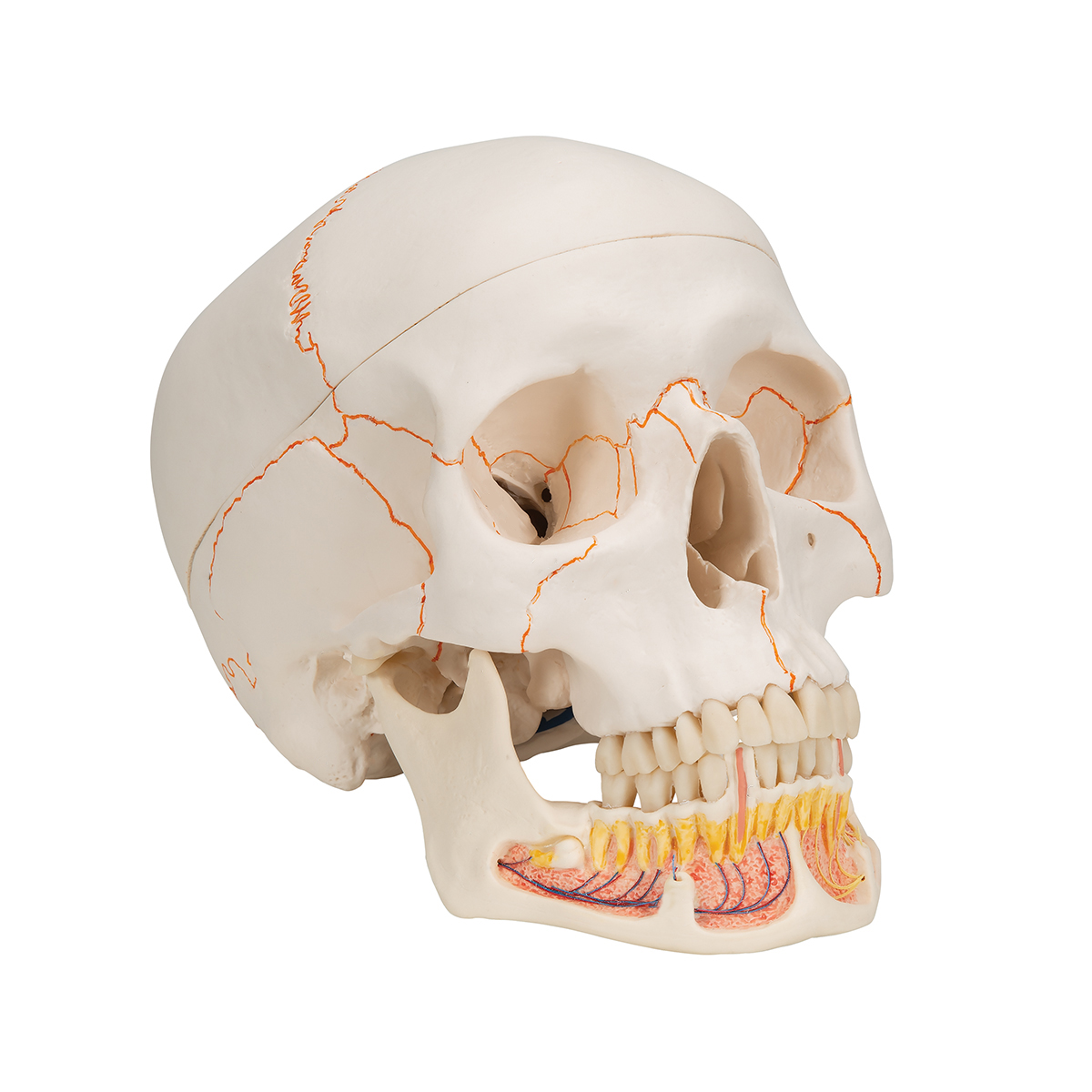
Learn about the anatomy of the skull bones and sutures as seen on ct images of the brain.
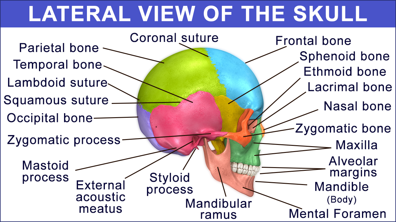
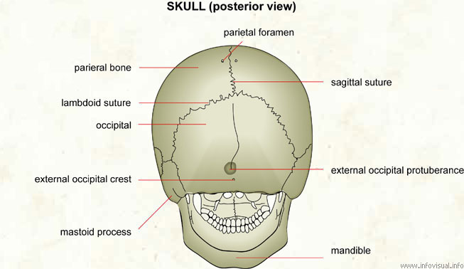
The simplest way to make the difference between the head and the face is to envision a ring that wraps around the head at the level the back of the head or occipital bone has four aesthetic bony regions. The frontal, parietal, temporal and occipital bones are joined at the cranial sutures. The occipital bone forms the back of the skull and the base of the cranium. The skull base is the inferior portion of the neurocranium. This anatomic region is complex and poses surgical challenges for otolaryngologists and neurosurgeons alike. The cranium and the mandible. In order to be light, the skull is made up by flat and irregular bones, and has hollow spaces called the sinuses. The anterior fossa is formed by the orbital plates of the frontal bone, cribriform plate of the ethmoid, and lesser wings of the sphenoid. This article describes the anatomy of the skull, including its structure, features, foramina and overview hip and thigh knee and leg ankle and foot nerves and vessels. Inside the skull, it forms the anterior cranial fossa, which contains the frontal lobes of the cerebrum. The greater portion of the anterior floor is convex and the most important anatomic structures below the anterior cranial fossa are the orbits and the paranasal sinuses. The major sutures are the coronal suture, sagittal suture, lambdoid suture and squamosal sutures. The skull performs vital functions.
The cranium and the mandible. Inside the skull, it forms the anterior cranial fossa, which contains the frontal lobes of the cerebrum. Excluding ear ossicles, it is made of 22 bones. The base of the skull (or skull base) forms the floor of the cranial cavity and separates the brain from the structures of the neck and face. Foramina inside the body of humans and other animals.
Human Skull Model | Plastic Skull Model | Dental Teaching ... from www.a3bs.com Learn more about the anatomy and function of the skull in humans and other vertebrates. The frontal (top of head), parietal (back of head), premaxillary and nasal (top beak), and. The skull has a single occipital condyle.7 the skull consists of five major bones: The skull performs vital functions. Human anatomy for muscle, reproductive, and skeleton. « back show on map ». Learn about the anatomy of the skull bones and sutures as seen on ct images of the brain. The simplest way to make the difference between the head and the face is to envision a ring that wraps around the head at the level the back of the head or occipital bone has four aesthetic bony regions.
Anatomy & physiology · anatomy and physiology.
So, the human skull consists of 23 bones. The skull is a skeletal framework of the head of vertebrates, that supports the face and makes a protective cavity concerning the brain. Anatomy & physiology · anatomy and physiology. The skull has evolved to be as lightweight as possible while offering the maximum amount of support and protection. The skull is the bony skeleton of the head. Inside the skull, it forms the anterior cranial fossa, which contains the frontal lobes of the cerebrum. « back show on map ». The axial & appendicular skeleton. It is comprised of many bones, formed by intramembranous ossification, which are joined together by sutures (fibrous joints). The skull base is the inferior portion of the neurocranium. Learn about the anatomy of the skull bones and sutures as seen on ct images of the brain. Upon reaching maturity, our skull bones fuse to produce a rigid protective shell for the soft nervous. The skull has a single occipital condyle.7 the skull consists of five major bones:
Anatomy & physiology · anatomy and physiology. The skull has a single occipital condyle.7 the skull consists of five major bones: Inferior view of base of the skull. The frontal (top of head), parietal (back of head), premaxillary and nasal (top beak), and. Anatomy next provides anatomy learning tools for students and teachers.
Skeletal System Diagrams from jb004.k12.sd.us Learn about the anatomy of the skull bones and sutures as seen on ct images of the brain. The skull bones can be classified into two groups: Human anatomy for muscle, reproductive, and skeleton. The skull begins to form prior to week 12 of embryogenesis. It supports and protects the face and the brain. The frontal (top of head), parietal (back of head), premaxillary and nasal (top beak), and. It is comprised of many bones, formed by intramembranous ossification, which are joined together by sutures (fibrous joints). The axial & appendicular skeleton.
It supports and protects the face and the brain.
• it has the supraorbital foramen, where the supraorbital the paired parietal bones make up the top and lateral aspects of the cranium. During childhood development, the skull bones remain somewhat separated, allowing for growth of the brain and skull. Human anatomy for muscle, reproductive, and skeleton. They don't move and united into a single unit. « back show on map ». 12 photos of the bone of back of skull. The bone is pierced by a large oval hole(the foramen magnum) through which runs the spinal cord. Learn about the anatomy of the skull bones and sutures as seen on ct images of the brain. Axial muscles of the head, neck, and back. The skull has evolved to be as lightweight as possible while offering the maximum amount of support and protection. William is a final year medical student in australia who has taught anatomy to tertiary science and. Excluding ear ossicles, it is made of 22 bones. Inferior view of base of the skull.


0 Komentar